
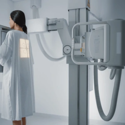
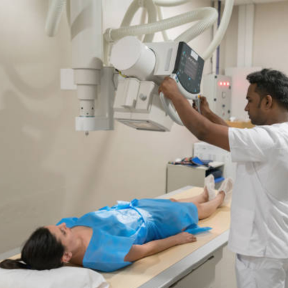
X-Ray
X-Ray is a non-invasive imaging technique used to visualize the internal structures of the body, especially bones and joints. It helps in diagnosing fractures, infections, abnormalities, and various medical conditions quickly and effectively.
The procedure involves passing a controlled amount of radiation through the body to capture images of internal organs or bones. It is commonly used in dental, orthopedic, and chest examinations for accurate diagnosis and treatment planning.
Benefits for Patients
- Quick and painless diagnostic process
- Accurate detection of fractures and conditions
- Essential for treatment planning
- Non-invasive and cost-effective
- Supports early diagnosis and intervention
Frequently Asked Questions
Learn more about X-Ray imaging and its role in modern diagnostics.
An X-Ray is a diagnostic imaging technique that uses low doses of radiation to produce images of the inside of the body, especially bones. It helps in detecting injuries, infections, and other conditions.
Yes, X-Rays are generally safe. The amount of radiation used is minimal and carefully controlled. Protective measures are used to ensure patient safety during the procedure.
X-Rays are recommended when a doctor needs to view bones, detect fractures, monitor conditions like arthritis, or examine the chest and lungs for infections or abnormalities.